cardiac conduction system pdf
Cardiac Conduction System⁚ A Comprehensive Overview
The cardiac conduction system is a network of specialized cardiac muscle cells that initiate and transmit the electrical impulses responsible for the coordinated contractions of each cardiac cycle. These special cells are able to generate an action potential on their own (self-excitation) and pass it on to other nearby cells (conduction), including cardiomyocytes.
Introduction
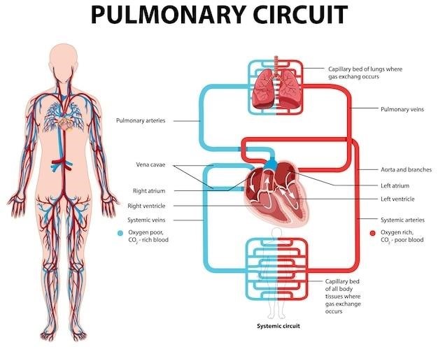
The human heart, a remarkable organ, tirelessly pumps blood throughout the body, ensuring the delivery of oxygen and nutrients while removing waste products. This vital function is orchestrated by a sophisticated electrical system known as the cardiac conduction system. Imagine the heart as a complex symphony, where each beat represents a harmonious interplay of electrical signals that control the rhythmic contractions of the heart muscle. This intricate network of specialized cells, distinct from the regular muscle cells of the heart, forms the foundation for the heart’s electrical activity.
The cardiac conduction system, often referred to as the heart’s electrical conduction system, is responsible for generating and transmitting electrical impulses that regulate the coordinated contractions of the heart. These impulses, like tiny electrical sparks, travel through a specific pathway within the heart, ensuring that the atria and ventricles contract in a synchronized manner, propelling blood efficiently throughout the circulatory system.
The Need for a Power Source
Consider the analogy of a central heating system. The pump, pipes, and radiators are essential components, but they are rendered useless without a power supply. The pump requires electricity to function, driving the circulation of heated water throughout the system. Similarly, the human heart, a remarkable pump, needs its own power source to operate. This power source is not a conventional electrical outlet, but rather a specialized network of cells within the heart itself, capable of generating and conducting electrical impulses.
The heart, like a finely tuned machine, relies on a steady stream of electrical signals to maintain its rhythmic contractions. These signals, originating within the heart’s own conduction system, act as the internal power source, ensuring that the heart beats in a coordinated and efficient manner. This intrinsic electrical system is vital for the heart’s ability to pump blood effectively, delivering oxygen and nutrients to every cell in the body.
Anatomy and Pathology of the Cardiac Conduction System
The cardiac conduction system, a marvel of biological design, is composed of specialized tissues that are distinct both histologically and electrophysiologically. These tissues are strategically located within the human heart, orchestrating its rhythmic contractions. Understanding the intricate anatomy and potential pathologies of this system is paramount for interventional electrophysiologists, enabling them to perform precise ablation procedures and implant pacemakers or defibrillators for the management of cardiac arrhythmias and heart failure.
The ability to accurately diagnose and treat these conditions relies heavily on a thorough grasp of the normal anatomy and function of the cardiac conduction system. Electrophysiologists must be able to identify the precise location of the conduction system’s components, as well as any abnormalities that may be present. This knowledge guides their interventions, ensuring effective and safe treatment for patients with heart rhythm disorders.
New Insights into Cardiac Anatomy of the Conduction System
The study of the cardiac conduction system has undergone a resurgence of interest in recent years, prompting a renewed focus on its clinical anatomy. This renewed attention is driven by a desire to fully appreciate the groundbreaking discoveries made over a century ago by Professor Sunao Tawara, a pioneer in the field of cardiac electrophysiology. Tawara’s meticulous research led to the identification of the atrioventricular (AV) node, a critical component of the conduction system, and laid the foundation for our current understanding of how electrical impulses travel through the heart.
By revisiting the clinical anatomy of the conduction system through the lens of modern clinical practice, researchers are gaining fresh insights into the intricate workings of this essential network. These insights are not only enhancing our understanding of normal cardiac function but also informing the development of more precise and effective treatments for a wide range of heart rhythm disorders.
The Role of the Cardiac Conduction System
The cardiac conduction system serves as the heart’s internal electrical wiring, orchestrating the rhythmic contractions that propel blood throughout the body. This specialized network of cells, distinct from the contractile muscle cells of the heart, initiates and propagates electrical impulses, ensuring that the atria and ventricles contract in a coordinated and synchronized manner. The system operates like a highly efficient electrical conductor, ensuring that the heart beats with a consistent rhythm and that blood is pumped effectively.
The cardiac conduction system’s role extends beyond simply initiating a heartbeat. It also plays a crucial role in regulating the heart’s rate and rhythm, adapting to changing demands on the body. For instance, during exercise or periods of stress, the conduction system speeds up the heart rate to deliver more oxygenated blood to the muscles; Conversely, during rest or sleep, the system slows the heart rate to conserve energy.
Components of the Cardiac Conduction System
The cardiac conduction system comprises a series of specialized cells, each with a unique role in generating and transmitting electrical impulses. These components work in concert to ensure a coordinated and efficient heartbeat.
The sinoatrial (SA) node, often referred to as the heart’s natural pacemaker, is located in the right atrium. It spontaneously generates electrical impulses, initiating the heartbeat. This electrical signal then spreads to the atria, causing them to contract. Next, the impulse reaches the atrioventricular (AV) node, situated between the atria and ventricles. This node acts as a gatekeeper, slowing down the electrical signal to allow the atria to fully contract before the ventricles begin.
The signal then travels through the bundle of His, a specialized pathway that connects the AV node to the ventricles. Finally, the impulse reaches the Purkinje fibers, a network of fibers that spread throughout the ventricles, stimulating them to contract. This coordinated contraction of the ventricles propels blood into the pulmonary and systemic circulation.
The Importance of the Cardiac Conduction System
The cardiac conduction system is paramount to the human heart’s ability to function effectively. It orchestrates the rhythmic contractions of the heart, ensuring that blood is efficiently pumped throughout the body. Without this intricate network of specialized cells, the heart would not be able to maintain a regular beat, leading to various cardiovascular issues.
The coordinated contractions generated by the cardiac conduction system are essential for maintaining adequate blood pressure and delivering oxygen and nutrients to the body’s tissues. Disruptions to this system, such as those caused by heart block or arrhythmias, can significantly impact cardiovascular health, potentially leading to fatigue, shortness of breath, dizziness, and even heart failure.
Understanding the workings of the cardiac conduction system is crucial for diagnosing and treating a wide range of heart conditions. Electrocardiograms (ECGs), for instance, provide insights into the electrical activity of the heart, allowing medical professionals to identify any abnormalities in the conduction system and tailor appropriate interventions.
The History of the Cardiac Conduction System
The journey of understanding the cardiac conduction system is a fascinating tale of scientific discovery and technological advancements. While the human heart has been a subject of curiosity for centuries, the precise mechanisms underlying its rhythmic beating remained a mystery until the late 19th and early 20th centuries.
In 1882, physiologist W.H. Gaskell coined the term “heart block,” describing a condition where electrical impulses are blocked from reaching the ventricles. This discovery marked a significant step towards understanding the conduction system’s role in heart function. The following year, Johannes E. von Purkinje described the ventricular conduction system, shedding light on the pathways through which electrical signals propagate in the heart.
However, it was Sunao Tawara, in 1906, who truly revolutionized our understanding of the cardiac conduction system. His groundbreaking work, “The Conduction System of the Heart,” provided the foundation for the anatomical and functional knowledge we have today. Tawara meticulously documented the atrioventricular node and its crucial role in regulating the flow of electrical signals from the atria to the ventricles.
The Anatomy of the Conduction System
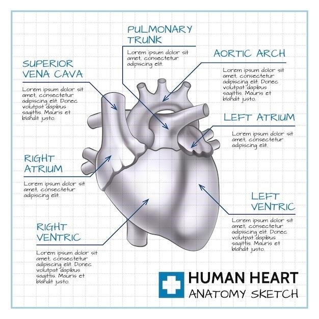
The cardiac conduction system, a complex network of specialized cells, acts as the heart’s electrical control center, orchestrating the rhythmic contractions that drive blood circulation. This system comprises four key components⁚ the sinoatrial (SA) node, the atrioventricular (AV) node, the bundle of His, and the Purkinje fibers.
The SA node, often referred to as the heart’s natural pacemaker, is located in the right atrium. It generates spontaneous electrical impulses that trigger atrial contraction. These impulses then travel to the AV node, situated between the atria and ventricles. The AV node acts as a gatekeeper, delaying the impulse slightly to allow the atria to fully contract before the ventricles begin.
From the AV node, the electrical signal travels through the bundle of His, a specialized pathway that divides into right and left bundle branches, extending down the septum between the ventricles. Finally, the signal reaches the Purkinje fibers, which spread throughout the ventricular walls, ensuring simultaneous and coordinated ventricular contraction.
The Electrocardiogram and the Cardiac Conduction System
The electrocardiogram (ECG), a widely used diagnostic tool in medicine, provides a visual representation of the electrical activity of the heart. It captures the electrical impulses generated by the cardiac conduction system, allowing healthcare professionals to assess the heart’s rhythm and identify any abnormalities. The ECG is a valuable tool for diagnosing various heart conditions, including arrhythmias, conduction defects, and myocardial ischemia.
The ECG waveform consists of distinct components, each representing a specific electrical event in the cardiac cycle. The P wave reflects atrial depolarization, initiated by the SA node. The QRS complex represents ventricular depolarization, as the electrical impulse travels through the ventricles. Finally, the T wave represents ventricular repolarization, as the ventricles return to their resting state. By analyzing these waveforms, healthcare professionals can identify specific patterns that indicate underlying heart problems.
The ECG is a non-invasive and relatively inexpensive test that plays a crucial role in understanding the function of the cardiac conduction system, aiding in the diagnosis and management of heart disease.
The Specialized Cardiomyocytes
The intricate workings of the cardiac conduction system rely on specialized cardiomyocytes, distinct from the contractile muscle cells that make up the bulk of the heart. These specialized cells possess unique properties that allow them to initiate and propagate electrical impulses, orchestrating the coordinated contractions that drive blood circulation.
These specialized cardiomyocytes are characterized by their ability to generate action potentials spontaneously, a property known as automaticity. This inherent rhythmicity is crucial for setting the pace of the heartbeat. The sinoatrial (SA) node, the heart’s primary pacemaker, is composed of specialized cardiomyocytes with the highest automaticity, initiating the electrical impulses that drive the cardiac cycle.
Furthermore, these specialized cardiomyocytes exhibit exceptional conductivity, allowing electrical impulses to travel rapidly and efficiently throughout the heart. This rapid conduction ensures that the heart contracts in a coordinated and synchronized manner, maximizing its pumping efficiency.
The specialized cardiomyocytes of the cardiac conduction system are essential for maintaining the heart’s rhythmic beat, ensuring the uninterrupted flow of blood throughout the body.
The Function of the Cardiac Conduction System
The cardiac conduction system serves as the heart’s electrical control center, ensuring that each beat is coordinated and efficient. This intricate network of specialized cells generates and transmits electrical impulses, orchestrating the synchronized contractions of the heart muscle, which propel blood through the circulatory system.
The process begins in the sinoatrial (SA) node, the heart’s natural pacemaker, located in the right atrium. Specialized cells within the SA node spontaneously generate electrical impulses, setting the rhythm for the heartbeat. These impulses then spread through the atria, causing them to contract and pump blood into the ventricles.
The electrical impulses then reach the atrioventricular (AV) node, a crucial junction between the atria and ventricles. Here, the impulses are briefly delayed, allowing the ventricles to fill with blood before contracting. This delay ensures that the atria and ventricles contract in a coordinated manner, maximizing the efficiency of blood pumping.
From the AV node, the electrical impulses travel through the bundle of His and its branches, reaching the Purkinje fibers, which extend throughout the ventricular walls. These fibers rapidly distribute the electrical signals, triggering the coordinated contraction of the ventricles, propelling blood out to the lungs and the rest of the body.
The Cardiac Conduction System⁚ A Closer Look
The cardiac conduction system is composed of specialized cells that are distinct from the contractile muscle cells of the heart. These cells are known as pacemaker cells and conducting cells, and they possess unique properties that enable them to generate and propagate electrical impulses. Pacemaker cells, located primarily in the sinoatrial (SA) node, have the ability to spontaneously generate electrical activity, initiating the heartbeat. These cells exhibit a rhythmic, spontaneous depolarization, which sets the intrinsic heart rate.
Conducting cells, found throughout the conduction system, are responsible for rapidly transmitting electrical impulses from one part of the heart to another. These cells are characterized by low resistance to electrical current, allowing for efficient conduction of the action potential. The unique structure and function of conducting cells enable them to efficiently transmit the electrical signal from the SA node to the ventricles, ensuring that the heart contracts in a coordinated and organized manner.
The specialized nature of these cells, along with their specific arrangement within the heart, allows for the precise control of heart rate and rhythm. The cardiac conduction system is a vital component of the cardiovascular system, ensuring that the heart pumps blood efficiently and effectively, delivering oxygen and nutrients to the body.
The Heart’s Electrical Conduction System
The heart’s electrical conduction system is a remarkable network of specialized cells that orchestrate the rhythmic beating of the heart. This intricate system acts as the heart’s internal pacemaker, generating and transmitting electrical impulses that trigger the coordinated contraction of the heart muscle. The system is comprised of specialized cells that are distinct from the contractile muscle cells of the heart, possessing unique properties that enable them to initiate and propagate electrical signals.
The sinoatrial (SA) node, often referred to as the “pacemaker” of the heart, is located in the right atrium. It is responsible for generating spontaneous electrical impulses at a regular rate, setting the rhythm for the heart’s contractions. These impulses travel through the atria, causing them to contract and pump blood into the ventricles.
The electrical signal then reaches the atrioventricular (AV) node, situated between the atria and ventricles. Here, the impulse is briefly delayed, allowing the ventricles to fill with blood before contracting. The signal then travels down the bundle of His, a specialized pathway that extends into the ventricles, and branches into the Purkinje fibers. These fibers distribute the electrical impulse throughout the ventricular muscle, triggering their coordinated contraction and expulsion of blood into the circulatory system.
The Importance of Understanding the Cardiac Conduction System
A comprehensive understanding of the cardiac conduction system is paramount for healthcare professionals, particularly those involved in the diagnosis and treatment of heart conditions. It provides a foundation for interpreting electrocardiograms (ECGs), the cornerstone of cardiac diagnostics, enabling the identification of abnormalities in the electrical activity of the heart. These abnormalities, such as heart blocks or arrhythmias, can be readily detected and analyzed based on the knowledge of the conduction system’s structure and function.
Furthermore, this knowledge is crucial for interventional electrophysiologists, who specialize in treating heart rhythm disorders. Understanding the anatomy and pathology of the conduction system allows them to perform safe ablation procedures, which involve the targeted destruction of abnormal electrical pathways, and to implant pacemakers or defibrillators to regulate heart rhythm. The insights gained from comprehending the conduction system are also essential for the development and refinement of new treatments for heart disease, including novel antiarrhythmic drugs and innovative device therapies.
Leave a Reply
You must be logged in to post a comment.